


3D printing saves dog’s skull
Researchers at a Canadian university used 3D printing technology to replace the majority of a dog’s cancer-ridden skull.
Michelle Oblak, DVM, DVSc, DACVS, veterinary surgical oncologist at the University of Guelph’s Ontario Veterinary College (OVC), performed the surgery and said she believes the procedure is the first of its kind in North America.
The patient, a nine-year-old dachshund named Patches, had a tumor, a multilobular osteochondrosarcoma that had grown so large it was weighing down her head, growing into her skull, and pushing dangerously close to her brain and eye socket.
Oblak performed the surgery at Cornell University’s College of Veterinary Medicine with a team that included Galina Hayes, BVSc, MRCVS, PhD, an assistant professor at Cornell and a former OVC colleague.
It was Hayes who first asked Oblak for advice on how best to treat the dog’s tumor.
Oblak was already doing research that involved using dogs as a disease model for cancer in humans. Her research centers on the use of digital rapid prototyping—creating product designs in 3D—for advance surgical planning. More specifically, creating 3D-printed implants for use in surgical reconstruction.
Patches’ tumor presented Oblak with both a challenge and a chance to test her research.
First, Oblak had to map the tumor’s location and size. Then, she worked with an expert in 3D printing, manufacturing, and design to create a 3D model of Patches’ head and tumor so she could “virtually” perform the surgery and see what would be left behind once the growth was removed.
Thanks to that 3D model, Oblak said, “I was able to do the surgery before I even walked into the operating room.”
Once Oblak determined the dimensions of the portion of skull she’d need to replace, she worked with a 3D medical printing company to adapt software designed for human medicine. Together, they created a skull plate to replace the part she planned to remove from Patches’ head.
That turned out to be about 70% of the top her skull.
Oblak said that typically, surgeries of this kind take a long time—once the portion of skull is removed, surgeons have to assess the damage and shape titanium mesh over the spot. In this case, no shaping was necessary: The 3D-printed plate fit into place perfectly. Oblak said Patches was asleep for about five hours, and “within half an hour after surgery, [she] was alert and looking around. It was amazing.”
Oblak said this technique will eliminate the need to model an implant in the operating room and reduce patient risk by shortening the time spent under anesthesia.
NEWStat asked Oblak what role 3D printing could play in veterinary hospitals.
“3D printing in companion animals has great potential for treatment but also for presurgical planning and teaching,” Oblak said. “I foresee that 3D anatomic models will start to be more available as soon as [open source] digital plans are available online and more people start to purchase the tabletop printers.”
“For presurgical planning, our group here has shown that 3D printing of skulls from a CT scan is incredibly accurate [and it’s] already used in many academic institutions,” Oblak said. “[And it’s] likely the use of 3D printing of bones for [surgical repair of] angular limb deformity, [and of] jaws and skulls for dental planning, will be more widespread in the very near future.”
What about 3D-printed custom implants for pets?
“From the perspective of custom implants, we are still a ways away from having easy accessibility and will likely always need to utilize service hubs for metal printing due to the cost of the printers,” Oblak said.
That day could be closer than we think, as the price of 3D printers continues to drop. Just seven years ago, a 3D printer could cost as much as $250,000.
Today, you could put one in your surgery suite for the price of a good laptop.
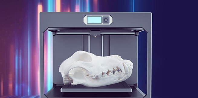
Photo credit: © iStock/Stefanie Garbutt/Sadie Lewandowski