Clinical
JAAHA: Treating a rare caudal mediastinal mass with nontraditional methods
A new case report in the Journal of the American Animal Hospital Association (JAAHA) outlines the nontraditional treatment of a rare caudal mediastinal mass.
Advertisement
A caudal mediastinal mass is a growth or tumor located in the lower part of the space in the chest between the lungs. In veterinary patients, such masses are rare, or at least not documented very often. These masses may be caused by several different factors, including bacteria, paraesophageal abscesses, or esophageal foreign bodies, among other things.
A new case report in the Journal of the American Animal Hospital Association (JAAHA) outlines the treatment of one of these rare cases. A Springer spaniel was brought into a veterinary teaching hospital, and a combination of CT imagery and surgical exploration revealed a golf ball-sized mass attached to the patient’s diaphragm and esophagus. Due to its location, the mass was deemed unresectable, so surgery was not an option.
Since surgical removal is the preferred method of dealing with these types of masses, the team had to figure out another way to treat the patient.
Read about what they found and ultimately did in the article, “Nonsurgical Management of a Caudal Mediastinal Granuloma,” in the latest issue of the Journal of the American Animal Hospital Association (JAAHA).
Disclaimer: Trends content is meant to inform, educate, and inspire by providing an array of diverse viewpoints. Therefore, any content published should not be viewed as an official stance, position, or endorsement by the American Animal Hospital Association (AAHA) or its Board of Directors.
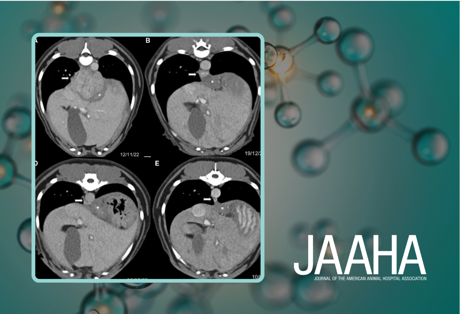
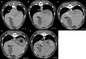
Photo credit: (A–E) Postcontrast soft tissue window computed tomography images at the level of the paraesophageal lesion (white arrow) showing a gradual decrease in size from November 2022 to May 2023. The esophagus (white asterisk) is distinguishable from the abscess. Photo courtesy of JAAHA
