Clinical
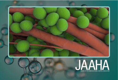
Gems from the Guidelines: Treating hypernatremia
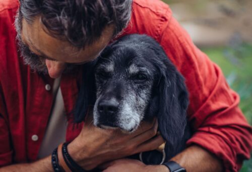
Summer is here, and that means fun in the sun–and the potential for heatstroke and the ingestion of seawater that can lead to dangerous levels of sodium in the blood.

Clinical
Summer is here, and that means fun in the sun–and the potential for heatstroke and the ingestion of seawater that can lead to dangerous levels of sodium in the blood.

Clinical

Clinical
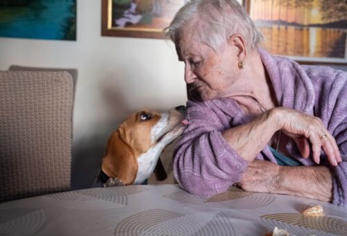
Community care
Culture and People

Clinical
By bringing AI-powered follow-up into the standard of care, not as a technology experiment, but as a clinical communication tool, veterinary practices can close the compliance gap that has persisted for decades.


Member Exclusive


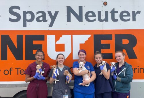
At veterinary schools across the nation, shelter medicine and community medicine programs are providing students with hands-on experience while expanding access to care for families in the area. It’s a winning combination that’s making a positive impact on young veterinarians and pet parents alike, and one that continues to grow and evolve.


AAHA Director Gregory Carastro, LVT, CVBL, talks about the importance of “taking care of the caregivers,” and building a workplace where employees can thrive and grow.