


Changes in fluid volume
Examples of common disorders causing changes in fluid volume
- Dehydration from any cause
- Heart disease
- Blood loss
Dehydration
Estimating the percent dehydration gives the clinician a guide in initial fluid volume needs; however, it must be considered an estimation only and can be grossly inaccurate due to comorbid conditions such as age and nutritional status (Table 5).
Fluid deficit calculation
Body weight (kg)3 % dehydration ¼ volume (L) to correct
General principles for fluid therapy to correct dehydration include the following:
- Add the deficit and ongoing losses to maintenance volumes. Replace ongoing losses within 2–3 hr of the loss, but replace deficit volumes over a longer time period. The typical goal is to restore euhydration within 24 hr (pending limitations of comorbid conditions such as heart disease).
- Frequency of monitoring will depend on the rate at which fluid resuscitation is being administered (usually q 15–60 min). Assess for euhydration, and avoid fluid overload through monitoring for improvement.
- Maintenance solutions low in Na should not be used to replace extracellular deficits (to correct dehydration) because that may lead to hyponatremia and hyperkalemia when those solutions are administered in large volumes.
TABLE 5
Dehydration assessment
* Not all animals will exhibit all signs.| Dehydration | Physical exam findings* |
| Euhydrated | Euhydrated (normal) |
| Mild (w ~ 5%) | Minimal loss of skin turgor, semidry mucous membranes, normal eye |
| Moderate (w ~ 8%) | Moderate loss of skin turgor, dry mucous membranes, weak rapid pulses, enophthalmos |
| Severe (. > 10%) | Considerable loss of skin turgor, severe enophthalmos, tachycardia, extremely dry mucous membranes, weak/thready pulses, hypotension, altered level of consciousness50 |
Hypovolemia
Hypovolemia refers to a decreased volume of fluid in the vascular system with or without whole body fluid depletion. Dehydration is the depletion of whole body fluid. Hypovolemia and dehydration are not mutually exclusive nor are they always linked. Hypotension may exist separately or along with hypovolemia and dehydration (Figure 1). Hypotension is discussed under “Fluids and Anesthesia.”
Common causes of hypovolemia include severe dehydration, rapid fluid loss (gastrointestinal losses, blood, polyuria), and vasodilation. Hypovolemic patients have signs of decreased tissue perfusion, such as abnormal mentation, mucous membrane color, capillary refill time, pulse quality, pulse rate, and/or cold extremity temperature.
Hypovolemia due to decreased oncotic pressure is suspected in patients that have a total protein , 35 g/L (3.5 g/dL) or albumin , 15 g/L (1.5 g/dL).19 Patients in shock may have hypovolemia, decreased BP, and increased lactate (. 2 mmol/L).20–22 Note that cats in hypovolemic shock may not be tachycardic.
Treating hypovolemia
When intravascular volume expansion without whole blood is needed, use crystalloids, colloids, or both. IV isotonic crystalloid fluids are the initial fluid of choice. If electrolytes such as K are needed in the emergent situation, administer through a second IV catheter. High K administration rates may lead to cardiac arrest; therefore, do not exceed 0.5 mmol/kg/hr.23–25
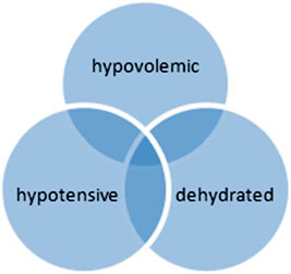
FIGURE 1
Patients may be hypovolemic, dehydrated, hypotensive, or a combination of all three.
How to administer crystalloids
- Standard crystalloid shock doses are essentially one complete blood volume.26
- Shock rates are 80–90 mL/kg IV in dogs and 50–55 mL/kg IV in cats.
- Begin by rapidly administering 25% of the calculated shock dose. Reassess the patient for the need to continue at each 25% dose increment.
- Monitor signs as described in the patient assessment portion of this document. In general, if 50% of the calculated shock volume of isotonic crystalloid has not caused sufficient improvement, consider either switching to or adding a colloid.
- Once shock is stabilized, replace initial calculated volume deficits over 6–8 hr depending on comorbidities such as renal function and cardiac disease.
When to administer colloids
- When it is difficult to administer sufficient volumes of fluids rapidly enough to resuscitate a patient and/or when achieving the greatest cardiovascular benefit with the least volume of infused fluids is desirable (e.g., large patient, emergency surgery, large fluid loss).
- In patients with large volume losses where crystalloids are not effectively improving or maintaining blood volume restoration.
- When increased tissue perfusion and O2 delivery is needed.27
- If edema develops prior to adequate blood volume restoration.
- When decreased oncotic pressure is suspected or when the total protein is ,< 35 g/L (or albumin is ,< 15 g/L).
- When there is a need for longer duration of effect. Preparations vary, and some colloids are longer lasting than crystalloids (up to 24 hr).28 Use of colloids can prolong the effects of hypertonic saline administration. The typical hydroxyethyl starch dose for the dog is up to 20 mL/kg/24 hr (divide into 5 mL/kg boluses and reassess). For the cat, the dose range is 10–20 mL/kg/24 hr (typically, 10 mL/kg in 2.5–3 mL/kg boluses).29–31 Titrate the amount of colloid infused to effect.
Simultaneously administering crystalloids and colloids
- Use this technique when it is necessary to both increase intravascular volume (via colloids) and replenish interstitial deficits (via crystalloids).
- Administer colloids at 5–10 mL/kg in the dog and 1–5 mL/kg in the cat. Administer the crystalloids at 40–45 mL/kg in the dog and 25–27 mL/kg in the cat, which is equivalent to approximately half the shock dose. Titrate to effect and continually reassess clinical parameters to adjust rate and type of fluid administered (crystalloid and/or colloid).
Using hypertonic saline
- To achieve the greatest cardiovascular benefit with the least volume of infused fluids (typically reserved for large patients or very large volume losses).
- To achieve translocation of fluids from the interstium to the intravascular space (e.g., for initial management of hemorrhage).
- In animals with hemorrhagic hypovolemic shock as a fastacting, low-volume resuscitation. Shock doses of hypertonic saline are 4–5 mL/kg for the dog and 2–4 mL/kg for the cat. Direct effects of hypertonic saline last 30–60 min in the vascular space before osmotic forces equilibrate between the intraand extravascular space. Once the patient is stabilized, continue with crystalloid therapy to replenish the interstitial fluid loss.
- In conjunction with synthetic colloids to potentiate the effects of the hypertonic saline.28,29
- Do not use hypertonic saline in cases of either hypernatremia or severe dehydration.
Treating hypovolemia due to blood loss
The decision of when to use blood products instead of balanced electrolyte solutions is based on the severity of estimated blood loss. Use of blood products is addressed elsewhere.32,33 If blood products are not deemed necessary, note that patients with low vascular volume (due to either vasodilation or hemorrhage) will benefit more from the use of colloids than crystalloids. Following 15 mL/kg of hemorrhage, even 75 mL/kg of crystalloid will not return blood volume to prehemorrhage levels because crystalloids are highly redistributed. Large volumes may be needed to achieve blood volume restoration goals, and large volumes may be detrimental to patients with normal whole body fluid volume but decreased vascular volume resulting from acute blood loss.34
Hypervolemia
Hypervolemia can be due to heart failure, renal failure, and/or iatrogenic fluid overload. Hypertension is not an indicator of hypervolemia. Treatment is directed at correcting underlying disease (e.g., chronic renal disease, heart disease), decreasing or stopping fluid administration, and (possibly) use of diuretics. Consider using hypotonic 0.45% sodium chloride as maintenance fluid therapy in patients susceptible to volume overload (such as those with heart disease) due to the decreased Na load.
Hyperthermia
Increased body temperature can rapidly lead to dehydration. Treatment includes administering IV replacement fluids while monitoring for overhydration. Subcutaneous fluids are not adequate to treat hyperthermia.